Record s20341
- Age: 77
- Sex: Female
Additional data
-
Comments
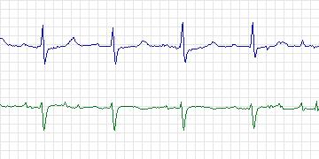
Lead 0: Two major ischemic episodes and several other less dramatic ischemic episodes characterized by down-sloping ST segments, generally associated with increases in heart rate. Lead 1: This lead, however, showed no evidence of ischemic ST changes. Electrode locations were not recorded. -
Diagnoses
  Coronary artery disease
  Hypertension
  Syncope
  Spinal stenosis
-
Treatment
-
Medication
Captopril Hydrochlorothiazide - Balloon Angioplasty: No
- Coronary Artery bypass Grafting: No
-
-
History
- Comments: Syncope
- Hypertension: Yes
- Left ventricular hypertrophy: Yes
- Cardiomyopathy: No
- Valve disease: No
- Electrolyte abnormalities: No
- Hypercapnia, anemia, hypotension, hyperventilation: No
- Atrioventricular nodal conduction delay: No
- Intraventricular conduction block: No
- Previous Myocardial Infarction: No
-
Previous tests
-
ECG stress test
- Date: No data
- Findings: Abnormal - infero-lateral ischemia, no angina
- Thallium/Stress echo: Positive antero-lateral ischemia
- Left ventricular function: Normal
-
Echocardiogram
Borderline left ventricular hypertrophy Ejection fraction > 55% - Coronary Arteriography: Done in 1998. Showed moderate 3-vessel disease, moderate left ventricular diastolic dysfunction, good ejection fraction
-
Baseline ECG
      Sinus bradycardia
      Left ventricular hypertrophy
      Left atrial abnormality
      Non-specific ST-T changes
-
-
Holter Recording
- Date: 12/11/1997
- Recorder: No data