Record s30661
- Age: 72
- Sex: Male
Additional data
-
Comments
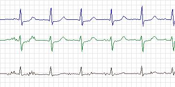
  This patient with extensive CAD shows a number of ST
  depressions associated with paroxysms of atrial
  tachycardia (PAT). Some ischemic changes were also
  seen in associations with increased heart rate which
  was not PAT. The patient has high-grade atrial ectopic
  activity with many bursts of PAT. Ischemia is most
  prominent in signal 0 (V4).
-
Diagnoses
  2-vessel coronary artery disease
  Unstable angina
  Previous coronary artery bypass grafting
  Left internal mammary artery graft to left anterior descending coronary artery
  Chronic obstructive pulmonary disease
  Hypertension
  Benign prostatic hypertrophy
-
Treatment
-
Medication
Aspirin Enalapril Metoprolol Prilosec Cardura Azmacort Ventolin Ipratropium - Balloon Angioplasty: No
- Coronary Artery bypass Grafting: Four grafts in 1995
-
-
History
- Comments: Unstable angina and coronary artery bypass grafting in 1995
- Hypertension: Yes
- Left ventricular hypertrophy: No
- Cardiomyopathy: No
- Valve disease: No
- Electrolyte abnormalities: No
- Hypercapnia, anemia, hypotension, hyperventilation: No
- Atrioventricular nodal conduction delay: No
- Intraventricular conduction block: No
- Previous Myocardial Infarction: No
-
Previous tests
-
ECG stress test
- Date: October, 1998
- Findings: 2 mm ST depression, stopped due to dyspnea
- Thallium/Stress echo: 1993 - no ischemia
- Left ventricular function: Normal - ejection fraction = 60%
- Echocardiogram: Normal left ventricular function
- Coronary Arteriography: 1992: lesions in left anterior descending coronary artery, right coronary artery disease
-
Baseline ECG
      Normal sinus rhythm
      Nonspecific ST changes
      Atrial premature beats
-
-
Holter Recording
- Date: 11/02/1999
- Recorder: Zymed