Record s30752
- Age: 79
- Sex: Male
Additional data
-
Comments
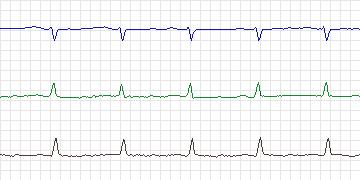
  This is the same patient as s30751. During this recording
  period there were a few minor ST depressions associated
  with increased heart rate. Only one event reached criteria
  for a significant episode, actually split into two episodes,
  in lead 0, and another one in lead 2.
- Diagnoses: Coronary artery disease
-
Treatment
-
Medication
Atenolol Simvastatin - Balloon Angioplasty: No data
- Coronary Artery bypass Grafting: Triple bypass in 1994
-
-
History
- Comments: Angina, coronary heart disease
- Hypertension: Yes
- Left ventricular hypertrophy: No
- Cardiomyopathy: No
- Valve disease: No
- Electrolyte abnormalities: No
- Hypercapnia, anemia, hypotension, hyperventilation: No
- Atrioventricular nodal conduction delay: No data
- Intraventricular conduction block: Left anterior fascicular block
- Previous Myocardial Infarction: No
-
Previous tests
-
ECG stress test
- Date: July, 1996
- Findings: Abnormal - 2 mm anterior ST segment depressions
- Thallium/Stress echo: Positive for inferior ischemia and wall motion abnormalities
- Left ventricular function: Hypokinesis and dyskinesis of infero-apical and septal walls, ejection fraction 44%
- Echocardiogram: Minimal left ventricular dysfunction & valvular regurgitation.
- Coronary Arteriography: Distal occlusion of posterior descending coronary artery
- Baseline ECG: Normal sinus rhythm, left anterior fascicular block, non-specific lateral ST-T abnormalities.
-
-
Holter Recording
- Date: 15/12/1999
- Recorder: Zymed