Record s30761
- Age: 72
- Sex: Female
Additional data
-
Comments
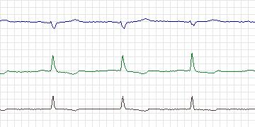
  Patient with coronary artery disease, and resting
  electrocardiogram is quite abnormal with ST down-sloping
  and biphasic T waves in leads 1 and 2. Ischemic episodes
  were present associated with increased heart rate. The
  changes were most convincing in lead 2 (with respect to
  ST morphology) but episodes were more frequent in lead 1.
-
Diagnoses
  Coronary artery disease
  Angina
  Hypertension
  Previous myocardial infarction
  Hypercholesterolemia
-
Treatment
-
Medication
Atenolol Imdur Cozar Lipitor Aspirin Lasix Synthroid (held during Holter) - Balloon Angioplasty: No
- Coronary Artery bypass Grafting: Yes
-
-
History
- Comments: Angina
- Hypertension: Yes
- Left ventricular hypertrophy: No data
- Cardiomyopathy: No data
- Valve disease: No data
- Electrolyte abnormalities: No data
- Hypercapnia, anemia, hypotension, hyperventilation: No data
- Atrioventricular nodal conduction delay: No
- Intraventricular conduction block: No
- Previous Myocardial Infarction: Yes, 1998
-
Previous tests
-
ECG stress test
- Date: No data
- Findings: Positive, infero-lateral ischemia
- Thallium/Stress echo: Ejection fraction 50%, inferior hypokinesis
- Left ventricular function: Abnormal - ejection fraction 40% - 50%
- Echocardiogram: No data
- Coronary Arteriography: Lesion in right coronary artery
- Baseline ECG: Sinus bradycardia at 55 bpm, infero-lateral ST depression and down-sloping.
-
-
Holter Recording
- Date: 22/11/1999
- Recorder: Zymed