Record s30771
- Age: 75
- Sex: Female
Additional data
-
Comments
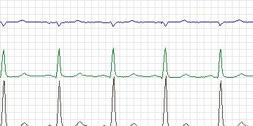
  The record shows ischemic ST depressions in all three
  leads which are associated with increased heart rate.
  In lead 2, during the first six and a half hours, there
  are also ST elevations recognized as rate-related
  episodes.
-
Diagnoses
  Coronary artery disease
  Previous myocardial infarction
-
Treatment
-
Medication
Atenolol Aspirin Pravachol - Balloon Angioplasty: No
- Coronary Artery bypass Grafting: No
-
-
History
- Comments: Coronary artery disease, old myocardial infarction
- Hypertension: No
- Left ventricular hypertrophy: No
- Cardiomyopathy: No data
- Valve disease: No data
- Electrolyte abnormalities: No data
- Hypercapnia, anemia, hypotension, hyperventilation: No data
- Atrioventricular nodal conduction delay: No
- Intraventricular conduction block: No
- Previous Myocardial Infarction: Yes, in 1997
-
Previous tests
-
ECG stress test
- Date: October, 1997
- Findings: Lateral precordial ST depression - positive test
- Thallium/Stress echo: Infero-apical dyskinesia with exercise.
- Left ventricular function: Ejection fraction 50%
- Echocardiogram: No data
- Coronary Arteriography: Lesions in left anterior descending coronary artery, right coronary artery, left circumflex coronary artery
- Baseline ECG: Normal sinus rhythm, non-specific ST-T changes in inferior leads and V6.
-
-
Holter Recording
- Date: 08/11/1999
- Recorder: Zymed