Record s30791
- Age: 74
- Sex: Male
Additional data
-
Comments
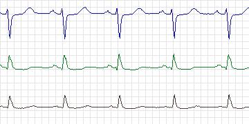
  The electrocardiogram at baseline is abnormal with
  ST concave depression. There also seems to be left
  axis deviation. The record shows numerous axis shifts.
  There are several episodes of ischemia related to
  increased heart rate, documented in all three leads.
  In the lead 0, however, the ST changes would be
  defined as rate-related if considered in isolation.
  They have been labeled as "ischemic" in light of
  the full 3-lead picture.
-
Diagnoses
  Coronary artery disease
  Hypertension
  Angina
-
Treatment
-
Medication
Atenolol Isosorbide (held during holter) - Balloon Angioplasty: No
- Coronary Artery bypass Grafting: No
-
-
History
- Comments: Angina, hypertension
- Hypertension: Yes
- Left ventricular hypertrophy: Yes
- Cardiomyopathy: No
- Valve disease: No
- Electrolyte abnormalities: No
- Hypercapnia, anemia, hypotension, hyperventilation: No
- Atrioventricular nodal conduction delay: No
- Intraventricular conduction block: No
- Previous Myocardial Infarction: No
-
Previous tests
-
ECG stress test
- Date: December, 1998
- Findings: Exercise induced ischemia. ST depression V4-V6
- Thallium/Stress echo: Positive blood pool scan (MUGA)
- Left ventricular function: Good - ejection fraction 60%
- Echocardiogram: No data
- Coronary Arteriography: Lesions in right coronary artery (100%), left anterior descending coronary artery (90%), left circumflex coronary artery (sequential 70%, 95%).
- Baseline ECG: Normal sinus rhythm, left ventricular hypertrophy
-
-
Holter Recording
- Date: 01/12/1999
- Recorder: Zymed